

Bone loss in the jaw is more than a dental inconvenience — it changes how your mouth functions and can subtly alter the shape of your face over time. Missing teeth, periodontal disease, and traumatic injury all remove the normal stimulation that keeps jawbone healthy and dense. Without treatment, the underlying bone can shrink significantly in the first year after tooth loss, making future restorative options more complicated and sometimes less predictable.
Jawbone provides the foundation for teeth and the soft tissues of the face. When bone resorbs, nearby teeth can shift, bite relationships can change, and the cheeks and lips may lose support, creating a sunken or aged appearance. These changes aren’t purely cosmetic — they affect how you chew, speak, and enjoy a comfortable bite.
Preserving or rebuilding lost bone restores the structural support that teeth and dental restorations depend on. For patients considering implants, bridges, or even well-fitting dentures, adequate bone volume is often the deciding factor in whether a given solution will be stable and long-lasting. Reconstructing the bone can also simplify future treatments and reduce the need for complex workarounds.
Understanding the causes of bone loss helps guide treatment. Active periodontal disease destroys the supporting bone around teeth, while the absence of a tooth eliminates the natural mechanical forces that maintain bone density. Early intervention and thoughtful planning give the best chance to protect both oral health and facial esthetics.
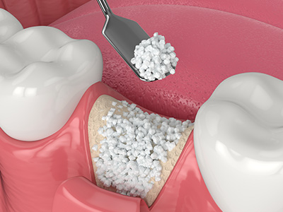
Bone grafting is a regenerative procedure that creates a supportive environment for your body to form new bone. Grafting material acts like a scaffold — it fills gaps, supports soft tissue, and provides a matrix that invites cells and blood vessels to grow in. Over time, the patient’s own bone gradually replaces the graft material and integrates with surrounding tissue.
There are several types of graft materials commonly used: bone taken from your own body (autograft), processed human donor bone (allograft), animal-derived bone that has been specially prepared (xenograft), and synthetic bone substitutes (alloplasts). Each has advantages: autografts are biologically active and integrate quickly, while donor and synthetic options avoid a second surgical site and still provide reliable support for regeneration.
Choosing the right material depends on the size of the defect, the patient’s overall health, and the intended restorative plan. Your clinician will explain the rationale behind a recommendation and how the chosen material supports predictable healing and long-term success.
One of the most effective strategies to reduce post-extraction bone loss is socket preservation — placing graft material into the tooth socket immediately after extraction. This approach minimizes the shrinkage that commonly happens in the months following tooth removal, keeping the ridge dimensions closer to their original contours and preserving options for future implants.
When a graft is placed later, after the site has already resorbed, the procedure typically involves a small incision to access the bone, placement of graft material, and closure of the tissues. These delayed grafts can still restore lost volume, but they may require more healing time or additional steps to prepare the area adequately for implant placement or other restorations.
Whether grafting occurs at the time of extraction or later, careful surgical technique and appropriate follow-up care are essential. The process includes protecting the graft from infection, controlling soft-tissue tension, and allowing adequate time for bone regeneration before placing implants or final restorations.
Grafting is not one-size-fits-all — certain areas of the mouth present unique challenges that call for specialized techniques. A common example is the upper back jaw, where the maxillary sinus can encroach on the available bone height. A sinus lift raises the sinus membrane and places graft material beneath it, creating the vertical dimension needed to support an implant without compromising sinus health.
Ridge augmentation focuses on rebuilding the horizontal contour of the jaw where bone volume has been lost. This procedure is often performed to restore the natural shape of the alveolar ridge for improved esthetics and to provide a stable foundation for crowns, bridges, or implant-supported teeth. In some restorative plans, both vertical and horizontal augmentation may be necessary to achieve a balanced, lasting result.
These techniques can be performed in a single stage or in multiple staged procedures depending on the anatomy and treatment goals. Advanced imaging and thorough treatment planning help determine the safest, most effective approach for each individual.
Protective measures are an important part of grafting protocols. Clinicians often use barrier membranes and biologically active substances to shield the graft from soft-tissue invasion while encouraging the migration of bone-forming cells. These adjunctive materials help the graft integrate more reliably and can shorten the overall timeline to a stable result.
Healing after grafting is gradual. Early weeks focus on soft-tissue recovery and the development of a blood supply, while the following months allow new bone to mature and strengthen. During this period, following postoperative instructions — such as avoiding pressure on the area and maintaining excellent oral hygiene — supports successful regeneration.
Complication rates are low when grafting is performed by experienced clinicians and when patients follow recommended care. Regular follow-up visits and appropriate imaging let your provider track the graft’s progress and time restorative steps, like implant placement, for when the new bone is ready to bear function.
At Advanced Dental Care of Las Colinas, our team approaches bone regeneration with thoughtful planning and evidence-based techniques to restore form and function. If you’re considering tooth replacement or worried about bone loss after extraction or periodontal disease, please contact us to learn more about your options and how a tailored grafting plan might preserve or rebuild the foundation your smile needs.
A bone graft is a surgical procedure that places bone or bone-like material where jawbone has been lost or is insufficient. The graft acts as a scaffold that encourages your body to grow new bone over time, restoring volume and strength. Dentists and oral surgeons use grafting to rebuild areas needed for function, aesthetics, or future restorative procedures.
Patients typically need a graft when bone loss follows tooth extraction, periodontal disease, trauma, or long-term tooth absence. Restoring bone can preserve facial contours, improve support for nearby teeth, and create a stable foundation for dental implants or bridgework. Your clinician will evaluate your situation with imaging and recommend grafting when it improves long-term outcomes.
Bone loss in the jaw undermines the support system for teeth and soft tissue, which can lead to tooth mobility, shifting, and additional tooth loss. Reduced bone height and width also change the way the lips and cheeks are supported, which may alter facial contours and make the lower face appear collapsed. These changes can affect chewing efficiency, speech, and overall oral function.
Preventing or treating bone loss helps protect remaining teeth and preserves the natural shape of the jaw and face. When bone is rebuilt through grafting, it restores support for prosthetic teeth and can improve comfort and function. Early assessment and intervention reduce the likelihood of progressive bone deterioration.
A bone graft can be placed at the time of tooth extraction or in a delayed fashion after the site has healed, and the timing depends on clinical factors. Immediate grafting at extraction preserves ridge width and height, minimizing the amount of bone that is lost in the first months after tooth removal. Delayed grafting is appropriate when infection, tissue condition, or other factors require healing before augmentation.
Your dentist or oral surgeon will assess the extraction site, overall oral health, and future restorative plans when recommending timing. Immediate grafting is often chosen to support future implants or preserve aesthetics, while staged approaches may be safer for sites that need additional healing or treatment. A personalized plan ensures the best balance of predictability and long-term stability.
Graft materials fall into several categories: autografts (from the patient), allografts (from human donors), xenografts (from animal sources), and synthetic bone substitutes. Autografts provide living bone cells and growth factors, while allografts and xenografts offer structural scaffolding without the need for a second surgical site. Synthetic materials can be engineered to resorb at controlled rates and support new bone formation.
The choice of material depends on the volume of augmentation needed, the site, patient preferences, and clinical goals. Your provider will weigh the pros and cons, such as healing potential, surgical complexity, and availability, to recommend the most appropriate option. In many cases, a combination of materials and biologic additives is used to enhance regeneration.
A sinus lift is a grafting procedure performed in the upper jaw beneath the sinus membrane to increase bone height for dental implants in the posterior maxilla. When the sinus floor is close to the alveolar ridge or bone has resorbed after tooth loss, there may not be enough vertical bone to securely anchor an implant. Lifting the sinus membrane and placing graft material creates the additional height needed for predictable implant placement.
Sinus lifts are recommended after careful imaging and planning to assess sinus anatomy and residual bone. Depending on the case, the graft may be placed at the time of implant surgery or in a separate stage to allow healing first. Your clinician will explain the risks, benefits, and expected timeline during treatment planning.
Bone grafting begins with a thorough clinical exam and imaging to map the defect and plan the approach. During the procedure the surgeon reflects the soft tissue, prepares the bone site, positions the graft material, and secures it as needed; membranes or biologic agents may be added to guide healing. The surgery can be performed using local anesthesia with or without sedation, depending on the complexity of the case and patient comfort needs.
Most graft procedures are outpatient and last from a short in-office procedure to a longer operation for extensive reconstruction. After placement the graft integrates gradually as your body replaces the scaffold with new bone, a process that can take several months. Your provider will give specific instructions on activity, diet, and medications to support healing and reduce the risk of complications.
Initial recovery from a bone graft usually involves some swelling, mild discomfort, and temporary changes in chewing or sensation near the surgical site. Pain and swelling are often managed with prescribed or over-the-counter medications, cold compresses, and short-term dietary modifications to avoid stressing the graft. It is important to follow activity restrictions and oral hygiene instructions to protect the graft during the early healing phase.
Your clinician will schedule follow-up visits to monitor integration and may use radiographs to document bone growth. Soft tissue healing typically completes in a few weeks, while new bone formation progresses over several months before any definitive restorative work like implant placement. Adhering to post-op care and attending scheduled appointments improves predictability and long-term success.
Yes, one of the primary goals of bone grafting is to create sufficient bone volume and quality to support dental implants. Grafts rebuild ridge width and height so implants can be placed in the ideal position for long-term function and aesthetics. Without adequate bone, implant stability and longevity are compromised, so grafting is often a necessary preparatory step.
The treatment plan connects grafting, healing time, and implant placement into a coordinated sequence tailored to each patient. After sufficient healing and imaging confirmation of new bone, implants can be placed with a higher likelihood of success. Your restoration team will discuss timing and the sequence that best fits your oral health goals.
Bone grafting is generally predictable, but like any surgical procedure it carries potential risks such as infection, wound dehiscence, graft failure, bleeding, or transient numbness in adjacent areas. Specific risks vary by graft type and location; for example, sinus lifts carry a small risk of sinus membrane perforation that may require repair. Your clinician will review individualized risk factors, medical history, and any medication considerations prior to treatment.
Careful surgical technique, appropriate material selection, and adherence to post-op instructions reduce complication rates. If a complication occurs, prompt evaluation and treatment help protect the graft and overall oral health. Open communication with your care team before and after surgery ensures concerns are addressed quickly.
At Advanced Dental Care of Las Colinas the team begins with a comprehensive assessment including digital imaging to evaluate bone volume, tooth position, and overall oral health. Treatment planning is individualized to align graft type, timing, and technique with each patient’s restorative and aesthetic goals. Where appropriate, the team coordinates care with implant specialists and uses modern biologic materials and membranes to enhance predictability.
Throughout treatment the practice emphasizes patient comfort, clear communication, and careful follow-up to monitor healing and outcomes. Sedation options and compassionate support are available for patients who need them, and the team provides detailed aftercare guidance to promote successful integration. Your provider will explain each step so you understand the plan and expected timeline before proceeding.
Quick Links