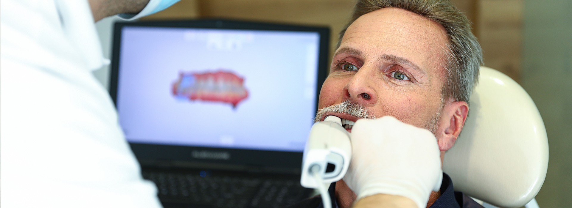


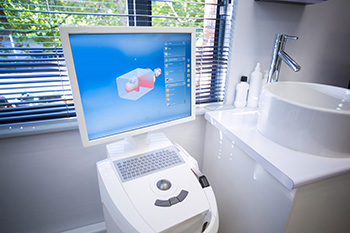
Digital impressions replace the traditional putty-and-tray method with a high-resolution optical scan of the teeth and surrounding tissues. Instead of filling a tray with viscous material and waiting for it to set, an intraoral scanner captures hundreds of thousands of individual data points as it passes over the dental surfaces. The result is a precise, three-dimensional model that can be viewed, manipulated, and shared instantly on a computer screen.
For patients, the difference is immediately noticeable: no unpleasant taste, no gagging, and no pressure from large impression trays. For clinicians, digital scans offer a repeatable and cleaner workflow that reduces the potential for distortion. The scanned data becomes a digital record that stays accessible for future treatment planning, communication with specialists, and tracking changes over time.
Although "digital impression" sounds technical, the goal is simple — to capture an accurate, usable replica of your mouth more comfortably and efficiently than traditional methods. This innovation has become a standard part of modern restorative and orthodontic care because it streamlines many steps that used to rely on manual laboratory work.
The scanning appointment is straightforward and efficient. After a brief examination, a clinician or trained team member will guide a compact wand-like scanner around your teeth and gums. The device uses a combination of visible light and advanced sensors to record surface geometry; a live image of your mouth appears on a monitor so you and the clinician can see the progress in real time.
Scans are completed in segments to ensure full coverage of crowns, preparation margins, and soft tissue contours. If saliva or blood obscures a small area, the clinician pauses briefly to gently control moisture and then resumes scanning. Because the process is visual and interactive, any missing or distorted areas can be corrected immediately rather than discovering an issue after a physical impression has been shipped to a lab.
Most patients describe the experience as quick and unobtrusive. The scanner’s software stitches captured frames into a seamless 3D model, and the clinician reviews the result on-screen to confirm margin visibility and occlusal relationships. When necessary, additional scans are taken within the same visit, eliminating the need for repeat appointments solely to correct impression problems.
Digital models deliver exceptional precision for crowns, bridges, inlays, and implant-supported restorations. Since the data is recorded directly from the mouth and processed by sophisticated algorithms, the likelihood of dimensional change that can occur with traditional impression materials is greatly reduced. This accuracy translates to better-fitting restorations and fewer adjustments chairside.
Another significant advantage is streamlined communication with dental laboratories and specialist providers. Digital files can be transmitted electronically in minutes, allowing lab technicians to begin design and fabrication without waiting for physical impressions to arrive by mail. When clinicians collaborate with specialists — for example, for implant planning or orthodontic aligners — the ability to share exact digital replicas enhances coordination and predictability.
Digital workflows also integrate with computer-aided design and manufacturing (CAD/CAM) systems, enabling precise milling or 3D printing of restorations and surgical guides. This level of integration reduces manual steps, which lowers the risk of human error and helps maintain consistent outcomes across complex treatment plans.
Because digital scans are captured and shared instantly, many restorative and orthodontic procedures move more quickly through the laboratory phases. For certain treatments, clinics that combine intraoral scanning with in-office milling can produce same-day ceramic crowns and onlays — a convenience that shortens treatment timelines and reduces the number of visits required. Even when lab fabrication is involved, electronic transfer expedites the process and can improve overall scheduling predictability.
For implant therapy and guided surgery, digital impressions are an essential component of modern planning. The digital model can be combined with CBCT data to fabricate surgical guides that support precise implant placement. For orthodontics, scanned models are used to plan tooth movement and fabricate clear aligners with tight tolerances, helping create consistent treatment progression.
The ability to store and compare digital scans over time also aids long-term care. Sequential scans make it easier to detect subtle changes in tooth position, wear patterns, or restorative margins, allowing clinicians to intervene earlier when adjustments or maintenance are needed.
From a patient perspective, the most obvious benefit is comfort. Eliminating tray-based impressions reduces gag reflex triggers and the unpleasant sensation of impression material in the mouth. For many patients — especially those with strong gag reflexes, anxiety, or sensitive oral tissues — digital impressions turn a potentially stressful part of treatment into a calm, manageable experience.
Digital scans also improve communication during consultations. Clinicians can rotate and zoom the 3D model on screen to show exact areas of concern, demonstrate proposed changes, and explain how a restoration or appliance will interface with nearby teeth. Seeing a visual representation of the problem and the intended solution helps patients make informed decisions and sets realistic expectations for outcomes.
Finally, digital records provide peace of mind. Accurate, retrievable scans reduce the likelihood of remakes and unnecessary appointments while creating a reliable archive that supports future treatments. Whether restoring a single tooth, designing a crown, or coordinating orthodontic care, the digital approach keeps patients better informed and more involved in their dental care.
At Advanced Dental Care of Las Colinas, we integrate intraoral scanning into our treatment workflows to deliver precise, patient-centered care. If you want to learn more about how digital impressions might change the way your dental work is planned and delivered, please contact us for more information.
Digital impressions are three-dimensional, computer-generated models of a patient’s teeth and surrounding oral tissues created with an intraoral optical scanner. These scans capture detailed surface geometry by collecting thousands of data points that are stitched together to form a precise digital replica. The resulting file replaces the need for traditional physical impressions made with impression material and trays.
Because the model is digital, it can be reviewed immediately on a monitor, adjusted if necessary, and transmitted electronically to a laboratory or used in chairside CAD/CAM workflows. Digital impressions support a range of restorative and orthodontic procedures and serve as a permanent, retrievable record for treatment planning. They reduce physical storage requirements and simplify long-term case management.
The process begins with the clinician using a small handheld intraoral scanner to gently move across the teeth and soft tissues while the device collects continuous images. Software stitches these images into a unified three-dimensional model in real time, allowing the dentist to verify margins, contact points, and overall scan quality during the appointment. If an area needs rescanning, it can be corrected immediately without remaking a physical impression.
Once the scan is complete, the digital file can be used for CAD/CAM design, sent electronically to a dental laboratory, or integrated with other digital records such as radiographs and treatment plans. This instant feedback loop shortens turnaround time and improves communication between the dental team and dental technicians. Many practices use compatible scanners such as the iTero scanner to ensure predictable, high-resolution captures.
Digital impressions are typically more comfortable than conventional putty impressions because they eliminate the need for impression trays and viscous materials that can cause gagging or discomfort. The scanning process is noninvasive and often faster, reducing time in the chair and the likelihood of repeat impressions. Patients also appreciate the ability to see their scan on a monitor, which helps them understand their condition and proposed treatment.
From a clinical perspective, digital impressions increase accuracy and reduce the incidence of remakes, which means fewer appointments and more predictable outcomes for patients. Electronic transmission of files speeds laboratory workflows, so restorations and appliances can be fabricated more efficiently. Many patients find the overall experience less stressful and more transparent than traditional impression techniques at Advanced Dental Care of Las Colinas.
Yes, high-quality digital impressions provide the level of accuracy required for crowns, bridges and implant restorations when captured and processed correctly. Modern scanners capture fine details of tooth margins and occlusal surfaces, and software tools allow clinicians to verify margins and contacts before finalizing the scan. Proper scanning technique, isolation of the field, and clinician experience are important factors in achieving optimal results.
For implant cases, digital impressions can be combined with implant libraries and virtual analogs to design precise prosthetics and surgical guides. Many laboratories accept or prefer digital files because they fit seamlessly into CAD/CAM workflows and reduce errors associated with physical model shipping or distortion. When a scan needs refinement, the digital workflow allows quick rescans rather than remakes from physical impressions.
Digital impressions are a key component of same-day restorative workflows that use in-office CAD/CAM systems to design and mill ceramic restorations. After capturing a digital impression, the restoration can be designed digitally, milled from a ceramic block, and delivered in a single visit when the practice has the appropriate CAD/CAM equipment and trained staff. This approach reduces total treatment time and minimizes the need for temporary restorations.
Not every case is suitable for same-day delivery; complex esthetic or multi-unit restorations may still require laboratory fabrication. However, many single-unit crowns, onlays and some veneers can be completed chairside using a fully digital workflow. The result is faster turnaround and fewer appointments for eligible patients without compromising material quality or fit.
Digital impressions reduce infection risk by eliminating the handling, storage and shipping of contaminated impression materials and stone models. Because the scan data is transmitted electronically, there is no need to transport physical impressions to an external laboratory, which lowers the number of contact points and surfaces that must be disinfected. The intraoral scanner tips are designed to be disinfected or used with disposable covers to maintain aseptic technique.
Electronic records also simplify sterilization protocols in the operatory since fewer physical components are exchanged between clinic and lab. This streamlined workflow supports modern infection-control standards and helps dental teams maintain a safer environment for patients and staff. Proper cleaning and disinfection of scanner components remain important to preserve equipment performance and patient safety.
Many patients who struggle with traditional impression trays find digital scanning more tolerable because it uses a slim wand rather than a full-size tray filled with impression material. The scanner can often be positioned to minimize contact with the soft palate and posterior tissues, reducing gag reflex triggers. For patients with limited mouth opening, experienced clinicians can use sectional scanning techniques to capture the necessary details in smaller passes.
That said, every patient is different, and some scans can be challenging due to anatomy, severe crowding, or patient movement. In those situations, clinicians adapt by using topical anesthetic, breaks during scanning, or alternative impression strategies as needed. The flexibility of digital workflows typically makes accommodating anxious or sensitive patients easier than with conventional methods.
After a digital scan, patients can usually return to normal activities immediately since the procedure is noninvasive and does not involve impression materials or temporary restorations in most cases. The dentist will review the scan, explain findings and discuss the proposed treatment plan while showing the three-dimensional images on a monitor. If a restoration is planned, the digital file will be sent to the CAD/CAM machine or to a laboratory for fabrication.
Patients may receive follow-up instructions related to the planned restoration or appliance rather than the scan itself. For example, if a crown or bridge is scheduled, the team will explain next steps, estimated timing and any temporary protections required while the final restoration is being fabricated. The digital record also allows easy retrieval for future visits or adjustments without repeating the scan unless anatomy has changed.
Digital impressions are the standard entry point for many clear-alignment systems because they produce precise three-dimensional models used to plan and sequence tooth movements. Scanned files can be uploaded to the aligner provider, where software simulates phased movements and produces custom aligners for each stage of treatment. This digital chain improves predictability and communication between the treating clinician and the aligner laboratory.
Using digital scans also simplifies monitoring progress and making midcourse corrections if needed, since new scans can be compared directly to initial models. For combined restorative and orthodontic cases, the digital workflow allows coordinated planning so restorative margins and tooth positions are optimized together. Many modern orthodontic and restorative systems rely on digital impressions for accurate, efficient treatment delivery.
Digital impressions play a central role in diagnosis, documentation and interdisciplinary treatment planning at the office of Advanced Dental Care of Las Colinas. Scans are integrated with radiographs and clinical photographs to create comprehensive records that inform restorative, implant and orthodontic decisions. The digital files make it simpler to collaborate with specialists and dental laboratories while maintaining a clear audit trail of treatment progress.
For long-term care, digital models allow clinicians to monitor changes in tooth wear, shifting, or prosthetic fit over time without repeated physical impressions. They also support efficient emergency workflows, remakes and replacements because digital files can be retrieved or updated quickly. This continuity of digital records benefits both clinical outcomes and patient convenience across multiple phases of care.
Quick Links