

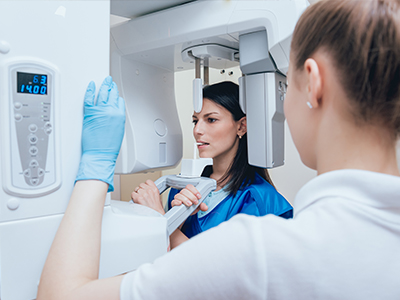
Digital radiography is a modern method for obtaining dental X-ray images using electronic sensors and computer systems instead of conventional film. When an image is taken, a small sensor placed in the mouth converts X-ray energy into a digital signal that is immediately processed by specialized software. That instant conversion eliminates the need for chemical development and produces a high-resolution image ready for review within seconds.
The captured images are stored directly in the patient’s electronic record, allowing clinicians to organize and retrieve them without handling physical film. Because the system records images as files, images can be enhanced, magnified, or measured with precision tools that help clinicians see details that might otherwise be missed. This digital format also makes it straightforward to create backups and maintain a robust image archive for long-term care.
Although the mechanics differ from film, the clinical goal remains the same: to reveal tooth, bone, and soft-tissue structures that cannot be seen during a visual exam. By converting X-rays into digital data, dental teams gain faster access to diagnostic information and the ability to present clear visual evidence during treatment planning discussions with patients.
One of the biggest advantages of digital radiography is the speed with which images become available for interpretation. Rather than waiting for film to be developed, clinicians can view images immediately and apply on-screen tools to adjust contrast, brightness, and zoom. These enhancements make subtle features — such as early decay, hairline fractures, or changes in bone density — easier to detect.
Digital sensors frequently produce higher-contrast images than traditional film, and software algorithms can sharpen details or suppress noise. This and the ability to compare current images with prior radiographs side-by-side help dentists monitor progression or healing over time with greater confidence. The result is a clearer, data-driven basis for diagnosis and treatment recommendations.
Because images can be reviewed instantly by multiple members of the clinical team, consultations are more efficient and collaborative. Specialists, hygienists, and restorative teams can all access the same files, which reduces miscommunication and supports coordinated care plans that are tailored to each patient’s needs.
Digital radiography significantly reduces radiation exposure compared with many conventional film systems. Modern sensors require less radiation to produce diagnostic-quality images, which is an important safety consideration for patients of all ages. Less exposure combined with precise targeting techniques helps clinicians maintain appropriate radiation stewardship while still obtaining the information needed for effective care.
From a comfort standpoint, sensors are thinner and more ergonomic than older film packets, making the imaging process less intrusive for many patients. The speed of image capture also shortens chair time, which reduces the number of positions and adjustments a patient must tolerate during a visit.
Because images appear on a monitor right away, clinicians can walk patients through what they see, improving understanding and trust. Visuals make it easier to explain diagnoses, compare “before and after” treatment expectations (when applicable), and discuss alternatives. This level of transparency helps patients make informed choices about their oral health without relying solely on verbal descriptions.
Digital radiography is often a central component of an integrated digital practice. Images can be imported into treatment-planning software, combined with intraoral scans, or paired with 3D imaging to create comprehensive views for complex procedures like implant placement or endodontic therapy. This interoperability streamlines planning and reduces the need for repeat exposures.
Because digital files are standardized, they can be shared securely with specialists, laboratories, or referring offices when collaborative care is required. Electronic transfer reduces the time and logistical friction associated with mailing physical films and supports faster decision-making. The ability to send precise images also helps ensure continuity of care when patients see multiple providers.
Integration with practice management software means images are linked to appointment notes, treatment plans, and billing records in a single patient file. That unified recordkeeping simplifies administrative tasks, minimizes data entry errors, and helps the clinical team focus on delivering care rather than managing paperwork.
Storing radiographic images digitally supports stronger recordkeeping practices. Files can be encrypted, access-controlled, and backed up according to professional and regulatory standards, protecting patient privacy while reducing the risk of lost or damaged records. Secure image management is an essential part of maintaining confidential and complete clinical histories.
Digital workflows also reduce environmental impact by removing the need for chemical developers, fixer solutions, and film waste. Eliminating these materials reduces hazardous waste and the associated handling requirements, aligning dental practices with greener operational practices.
Finally, having a consistent, searchable image archive enhances long-term patient care. Clinicians can quickly review historical images when evaluating changes, making it easier to detect trends and respond proactively to emerging concerns. That continuity contributes to better outcomes and more efficient, patient-centered care.
In summary, digital radiography modernizes how dental teams capture, view, and manage X-ray images. It delivers faster results, improved diagnostic detail, enhanced patient communication, and streamlined integration with other digital tools used in contemporary dentistry. If you’d like to learn more about how Advanced Dental Care of Las Colinas uses digital imaging in patient care, please contact us for additional information.
Digital radiography uses electronic sensors and computer software to capture, view, store, and manage dental X-ray images. Instead of traditional film, a small sensor records the image and transmits it instantly to a computer for review. This technology supports clearer images, faster workflows, and easier long-term record keeping.
Because images are stored digitally, they can be enhanced, measured, and compared over time without physical film degradation. The process eliminates film processing chemicals and reduces handling, which is better for the environment. Digital files also make it simpler to include radiographs in a patient’s electronic health record for coordinated care.
Digital radiography captures images using electronic sensors rather than radiographic film, which removes the need for chemical development. Images are available instantly on a monitor and can be adjusted for contrast, brightness, and magnification to aid diagnosis. These enhancements often reveal details that are harder to see on conventional film.
Digital systems typically require less radiation to produce diagnostic images compared with older film-based techniques. Digital images are also easier to store, duplicate, and transmit securely to other clinicians when consultation or referral is needed. The combination of improved image manipulation and streamlined sharing supports faster, more accurate treatment planning.
Digital radiography is considered safe and is designed to minimize radiation exposure while providing clinically useful images. Advances in sensor sensitivity and image processing mean many exams can be completed using lower doses than traditional film X-rays. Dental teams follow established safety protocols such as proper positioning and the use of protective shielding when appropriate.
Equipment is maintained and calibrated to ensure consistent performance and patient safety. Clinicians only take radiographs when they are clinically justified based on symptoms, examination findings, or monitoring needs. If you have specific concerns about exposure, your dental team can explain why a particular image is recommended and how risk is managed.
A digital dental X-ray begins with placement of a small sensor inside the mouth or positioning an external sensor for panoramic or extraoral images. The sensor is connected to the computer system and records the X-ray exposure, which is then converted into a digital image. The whole capture process typically takes only a few seconds for each image.
Once captured, the image appears on a monitor immediately for the clinician to review and adjust if needed. If an image is unclear, a quick retake is possible with minimal additional exposure. The digital workflow streamlines record keeping and helps the clinician explain findings to the patient using on-screen visuals.
Digital X-rays provide sharp, high-contrast images that help clinicians detect cavities, bone loss, root issues, and abnormalities that may not be visible during a clinical exam. Image enhancement tools such as magnification and contrast adjustment improve the clinician’s ability to evaluate fine details. Sequential images can be compared over time to monitor disease progression or healing.
Because images are stored electronically, they can be integrated into treatment planning software for implant placement, orthodontic assessment, and surgical preparation. Sharing clear digital images with specialists supports collaborative care and informed decision making. Overall, digital radiography enhances diagnostic accuracy and helps clinicians communicate treatment options more clearly to patients.
Yes. One of the advantages of digital radiography is that images can be exported and shared electronically with other clinicians when referrals or second opinions are needed. Digital files can be sent securely or provided on request so that specialists have the same high-quality images for review. This capability improves coordination of care and reduces the need for duplicate imaging when records are transferred properly.
Sharing is performed in accordance with privacy and security standards to protect patient information. The digital format also allows the practice to include radiographs in a comprehensive electronic health record for streamlined access by authorized providers. Patients are encouraged to bring any recent external imaging or share details of prior care to support continuity.
Dental radiographs are generally avoided during pregnancy unless the information gained will directly affect urgent dental care. When radiographs are necessary, clinicians take extra precautions such as using a lead apron and thyroid collar and limiting the number of exposures. Modern digital systems also reduce the radiation dose compared with older film techniques, which further decreases risk.
Decisions about imaging during pregnancy are made on a case-by-case basis, balancing the diagnostic benefit with the need to minimize exposure. If you are pregnant or suspect you may be, inform the dental team before any X-rays are taken so staff can apply appropriate safety measures and discuss alternatives. Coordination with your medical provider can help ensure the best approach for both dental and prenatal health.
Digital radiography is commonly used for pediatric patients because sensors and exposure settings can be adapted to the child’s size and clinical needs. Lower exposure settings and modern sensor sensitivity make it possible to obtain diagnostic images with minimal radiation. Pediatric imaging protocols also emphasize judicious use of X-rays, taking only those images necessary for accurate diagnosis and treatment planning.
Dental teams experienced with children use positioning aids and clear instructions to help the child remain still and comfortable during the procedure. Frequency of imaging follows clinical guidelines and is based on individual risk factors, development, and symptoms. Parents should discuss any concerns with the clinician so the imaging plan aligns with the child’s needs and safety considerations.
At Advanced Dental Care of Las Colinas, digital radiography is integrated into routine exams and treatment workflows to support accurate diagnoses and individualized care plans. Digital images allow the team to demonstrate findings visually, explain treatment options, and document progress across visits. The ability to enhance and compare images helps clinicians monitor healing and the long-term stability of restorations and implants.
Storing radiographs electronically improves record keeping and coordination with specialists when collaborative care is required. The practice follows best-practice safety protocols and maintains equipment to ensure reliable image quality. Patients benefit from quicker appointments, clearer communication, and more informed clinical decisions supported by modern imaging technology.
No special preparation is usually required for routine dental radiographs, but there are a few common precautions to follow. Wear comfortable clothing and avoid wearing jewelry or removable metal objects that could interfere with imaging. If you are pregnant, think you may be pregnant, or have specific health concerns, inform the dental team before the appointment so appropriate measures can be taken.
If you have recent radiographs from another office, bringing copies or arranging for electronic transfer can prevent unnecessary repeat imaging. Also let the clinician know about any previous adverse reactions to imaging procedures or implants that could affect positioning. Clear communication with your dental team helps ensure the imaging visit is efficient, safe, and focused on your diagnostic needs.
Quick Links