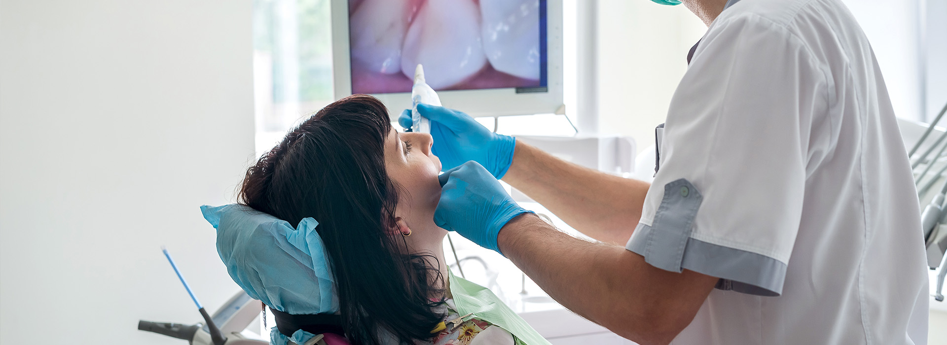

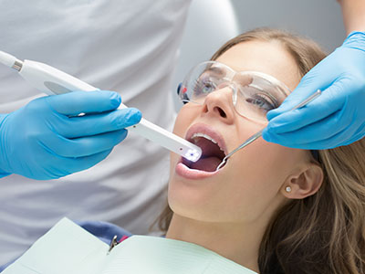
An intraoral camera is a compact, pen-sized imaging device designed to capture detailed, full-color views of the inside of the mouth. Unlike traditional inspection with a mirror and light alone, this tool delivers high-resolution images of teeth, gums, and other oral tissues so clinicians can see small problems that might otherwise be missed. The camera's close-up perspective makes it easier to identify early signs of decay, hairline fractures, worn restorations, and changes in soft tissue color or texture.
Because the camera records what the clinician sees in real time, it transforms routine visual exams into a documented part of your dental record. These images offer a visual baseline that clinicians can compare over time to track progression or healing. For patients, seeing a clear image on-screen turns abstract descriptions into concrete information — a powerful aid for understanding oral health status.
In short, the intraoral camera extends the clinician's vision into areas that are difficult to inspect with the naked eye, improving diagnostic confidence. With precise imaging, dentists can make more informed decisions about preventive care and when restorative work is necessary, leading to more conservative and targeted treatments.
One of the most immediate benefits of intraoral imaging is improved communication. When clinicians can show patients exactly what they are seeing, conversations about treatment options become clearer and more productive. Patients are better able to weigh conservative monitoring against restorative care because they can visually grasp the nature and extent of an issue rather than relying solely on verbal explanations.
For dental teams, these images help streamline treatment planning. Clear photos and video clips can be annotated, magnified, or paused to focus on areas of interest during consultation. This shared visual reference reduces misunderstandings, aligns expectations, and helps patients feel more actively involved in decisions about their care.
At Advanced Dental Care of Las Colinas, intraoral imaging is integrated into the exam workflow so that communication is consistent and patient-centered. Using these images during reviews and consent discussions supports transparency and builds trust — patients leave appointments with a clearer sense of what was found and why a particular recommendation is being made.
Modern intraoral cameras produce images that are easily stored in digital patient records. These files become part of the clinical history and can be retrieved for future comparisons, insurance documentation, or referral purposes. High-quality images reduce ambiguity and ensure that specialists, labs, and other providers see precisely the same anatomy and problem areas when collaborating on a case.
Sharing digital images with a laboratory or an oral surgeon improves predictability for complex treatments like crowns, implants, or periodontal procedures. When outside providers receive clear, annotated images, they can plan restorations or interventions more accurately and prepare the necessary instruments or materials in advance, which often shortens treatment timelines and reduces unexpected adjustments.
Because these images are date-stamped and retained in the record, they also help with long-term monitoring. Clinicians can measure changes, reassess treatment outcomes, and document healing in a way that numerical notes alone cannot. This continuity of information supports both preventive strategies and restorative care over the years.
From a patient perspective, intraoral cameras are comfortable and noninvasive. The slender housings and ergonomic tips are designed to move smoothly within the mouth, allowing clinicians to capture useful angles without discomfort. For patients who feel anxious about dental exams, being able to watch the process on-screen and understand what is happening often reduces fear and increases cooperation during appointments.
Safety is an inherent part of using intraoral cameras in a clinical setting. Most units have removable sleeves or barriers that are changed between patients, and the devices are handled according to infection-control protocols. When combined with standard sterilization and hygiene practices, intraoral imaging is a safe adjunct to examination and documentation.
Beyond comfort and safety, these images provide reassurance. Seeing a problem early — or seeing that an area is healthy — helps patients prioritize care and avoid surprises. This clarity supports better oral-health decisions and encourages adherence to recommended preventive or restorative plans.
Not all intraoral cameras are created equal. Contemporary devices deliver higher resolution, improved color fidelity, and enhanced lighting that reduce glare and shadowing. Many models offer autofocus, adjustable light intensity, and small-diameter heads that facilitate access to posterior teeth and subgingival margins. These technical advances translate directly into clearer images and more reliable clinical interpretation.
Integration with practice software is another hallmark of modern systems. Seamless transfer of images into electronic health records, the ability to annotate or crop photos, and connectivity for secure sharing are essential features that make these cameras valuable everyday tools. Reliable backup and indexing mean clinicians can pull up past images in seconds during follow-up visits.
Ongoing staff training and proper device maintenance also influence image quality. Clinicians and assistants who know how to position the camera, optimize lighting, and capture the right angles will consistently produce diagnostically useful images. Regular calibration and adherence to manufacturer guidelines keep the equipment operating at peak performance.
Intraoral cameras have become a standard part of modern dental care because they strengthen diagnostics, improve communication, and create a visual record that supports long-term treatment planning. If you would like to learn more about how this technology is used during exams or how images are incorporated into your care, Advanced Dental Care of Las Colinas is happy to explain the process and answer your questions. Please contact us for more information.
An intraoral camera is a small, pen-sized digital camera designed to capture high-resolution, full-color images inside the mouth. It provides real-time video and still images of teeth, gums, and other oral tissues so clinicians and patients can view detailed close-ups on a monitor. This technology helps reveal surface features that can be difficult to see with the naked eye, such as hairline cracks, worn restorations, and early soft-tissue changes.
The device is handheld and typically fitted with a disposable sleeve for infection control, allowing quick, noninvasive imaging during routine exams. Images can be paused, enlarged, and saved for documentation or comparison over time. Because the output is visual and immediate, it serves as a practical diagnostic and patient-education tool in modern dental practice.
During an exam, the dentist or hygienist gently positions the intraoral camera inside the mouth to capture views of individual teeth and surrounding tissues. The clinician can rotate the camera to inspect occlusal surfaces, interproximal areas, and the gum line while the images display on a chairside monitor. This live view allows the clinician to point out specific concerns and to take still photos for the patient record.
Captured images are often saved directly into the patient’s digital file for future reference and for treatment planning. These saved photos can be reviewed during follow-up visits to track changes or to show before-and-after progress of therapies. The process is fast and typically adds only a few minutes to a routine appointment.
An intraoral camera gives patients a clear, visual explanation of oral health conditions that are otherwise difficult to observe. Seeing a close-up image of a problem area helps patients understand diagnoses and compare treatment options more easily than verbal descriptions alone. This transparency supports informed decision making and improves communication between the care team and the patient.
In addition to education, intraoral images improve documentation and continuity of care by creating a precise visual record. Those images can be shared with specialists, dental laboratories, or retained for monitoring healing and treatment outcomes. Overall, the technology enhances trust and helps patients participate actively in their dental care.
Yes, intraoral cameras are safe and designed for patient comfort; they are noninvasive and cause little to no discomfort. The camera tip is small and smooth, and clinicians use disposable protective sleeves to maintain infection control and hygiene. Most patients experience only minimal awareness of the device while clinicians capture the necessary views.
Because imaging is quick, clinicians can pause or reposition the camera if a patient needs a break or experiences sensitivity. Staff are trained to work gently and efficiently, and they can adapt technique for patients with limited mouth opening or sensitivity issues. Overall, the procedure is well tolerated by the vast majority of patients.
Images taken with an intraoral camera are saved directly into the patient’s secure digital record so they become part of the clinical chart. These files are organized and timestamped for easy retrieval during future visits and for longitudinal comparison. Storing images digitally streamlines documentation and helps ensure accurate clinical records over time.
With the patient’s consent, clinicians can export or electronically share images with other dental specialists, dental laboratories, or outside providers to support coordinated care. Advanced Dental Care of Las Colinas uses these shared images to collaborate when referrals or lab work are required, improving treatment planning and communication across the care team.
An intraoral camera is effective at revealing visible signs of decay, cracks, worn fillings, and soft-tissue conditions on tooth and gum surfaces. It excels at highlighting surface irregularities and early enamel changes that merit closer examination. However, because it captures only surface images, it cannot visualize internal tooth structure or bone that may be revealed on X-rays.
For comprehensive diagnosis, clinicians combine intraoral imaging with other diagnostic tools such as digital radiography and clinical probing. Together, these methods provide a fuller picture of oral health so the dentist can confirm a diagnosis and recommend the most appropriate course of treatment.
An intraoral camera brings visual clarity to conversations about oral health by showing patients the same images the clinician sees. This shared perspective makes it easier to explain findings, demonstrate problem areas, and discuss why a recommended treatment is necessary. Visual evidence often helps patients better understand the urgency and expected outcomes of suggested care.
The camera also supports collaborative treatment planning by allowing patients to ask focused questions while the clinician points to specific images. Saved photos can be reviewed later or included in pre-treatment consultations, which helps patients feel more informed and involved in decisions about their care.
An intraoral camera captures color images of tooth surfaces and soft tissues, providing detailed photographic views of what is visible inside the mouth. Digital X-rays, by contrast, use radiation to image internal structures such as tooth roots, interproximal decay beneath the enamel, and supporting bone. Each tool reveals different types of information and neither replaces the other for a complete assessment.
Clinicians commonly use both technologies together: intraoral photography for visual surface assessment and education, and digital radiography for evaluating internal anatomy and bone health. Combining these diagnostic tools improves accuracy and helps the dental team develop comprehensive treatment plans.
Yes, intraoral cameras can be adapted for children and patients prone to gagging by using pediatric-sized tips and gentle technique. Clinicians shorten imaging sessions, introduce the device slowly, and use distraction or reassurance to help younger or anxious patients remain comfortable. The small, smooth camera heads and quick captures reduce the chance of triggering a gag reflex compared with larger instruments.
If a patient experiences difficulty, the dental team may take images in stages, adjust positioning, or use alternative imaging angles to obtain the necessary views. Experienced staff are skilled at modifying their approach to accommodate a wide range of patient needs while still collecting useful diagnostic images.
Intraoral images are incorporated into treatment planning by documenting the initial condition and supporting visual explanations of proposed procedures. Clinicians reference these photos when outlining treatment steps, discussing options, or coordinating care with specialists and dental laboratories. The images help ensure that everyone involved has the same clear visual information guiding decisions.
During follow-up visits, saved photos allow clinicians to compare progress, confirm expected healing, and assess the success of restorations or other therapies. Advanced Dental Care of Las Colinas uses intraoral imaging routinely to track outcomes and maintain accurate, visual records that support continuity of care for every patient.
Quick Links