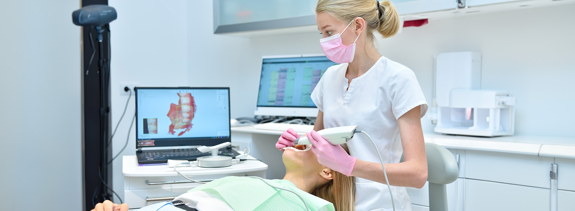

The iTero® Scanner has transformed how modern dental teams capture the precise geometry of teeth and bite relationships. By replacing traditional impression materials with a guided, digital scanning process, the iTero® Scanner provides a high-resolution, three-dimensional view of the mouth that clinicians can use immediately for diagnostics, treatment planning, and appliance fabrication. This page explains how the technology works, why it matters for outcomes, and how our team leverages it to make visits more predictable and comfortable.
The iTero® Scanner is an intraoral wand that collects a rapid series of images and stitches them together into a single, accurate 3D model. During a scan, the clinician moves the wand around the dental arches while the software guides the capture sequence, ensuring full coverage of teeth, soft tissues, and occlusal contacts. The result is a digital impression that shows fine surface detail, tooth position, and how the upper and lower teeth meet — information that’s essential for restorative and orthodontic care.
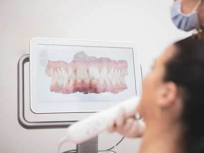
Scans are processed in real time, so clinicians can review the model with patients immediately after capture. This instant feedback speeds clinical decision-making; adjustments can be made on the spot if any area needs rescanning, reducing uncertainty and eliminating the need for remakes. The scanned data can be viewed from multiple angles, zoomed and annotated, which helps illustrate conditions and proposed treatments in a way that’s easy for patients to understand.
Because the iTero® Scanner produces a digital file rather than a physical cast, it becomes a versatile asset in the patient record. It can be archived for monitoring changes over time, compared against future scans, or securely transmitted to dental laboratories and specialty partners. That digital workflow creates efficiencies that benefit both the practice and the patient through faster turnarounds and more precise appliance fabrication.
Traditional impression techniques are subject to material distortion, handling variability, and human error, all of which can lead to poorly fitting restorations or appliances. The iTero® Scanner minimizes those sources of variation by capturing hundreds of micro-images and aligning them algorithmically to produce a highly accurate 3D model. That consistency improves the fit of crowns, bridges, veneers, and orthodontic aligners, reducing the need for adjustments or redo work.
Clinical predictability comes from both the level of detail and the reproducibility of scans. Because digital captures are standardized, clinicians can rely on measurements taken from the model for margin design, occlusal assessment, and treatment planning with greater confidence. Laboratories and digital mills receive a precise, machinable file, which increases the chance that the final prosthesis will match the intended contours, contacts, and occlusion.
This precision also supports interdisciplinary cases. When restorative, implant, and orthodontic specialists collaborate, having a single, accurate digital dataset avoids miscommunication and helps coordinate complex care. The iTero® Scanner’s output integrates cleanly with planning software and digital labs, so every member of the care team can work from the same, reliable information.
For many patients, the most immediate benefit of digital scanning is comfort. Conventional impressions often require bulky trays and impression material that can trigger gagging, taste sensitivity, or general discomfort. Scanning replaces that experience with a small, ergonomic wand that glides along the teeth; most patients describe the session as quick and unobtrusive. Because the scan is non-invasive and performed without putting material into the mouth for extended periods, patients with sensitive gag reflexes or anatomical challenges tend to prefer this method.
The speed of a digital workflow also reduces chair time. Scans typically take only a few minutes, and the digital file is immediately available for review and transmission. For procedures that require lab-fabricated components, this can shorten the total treatment timeline because the lab receives a ready-to-use digital file without waiting for physical cast shipment, disinfection, and manual scanning.
Another practical advantage is predictability on future visits. Digital archives allow clinicians to compare past and present scans side-by-side, tracking wear, movement, or tissue response over months and years. That continuity improves preventive care and makes it easier to spot subtle changes early, offering patients a more proactive approach to oral health management.
The iTero® Scanner is designed to work with contemporary digital dentistry systems, so files can be exported to CAD/CAM mills, 3D printers, and certified laboratories. For restorative dentistry, that means precise crowns, inlays, onlays, and bridges that fit more predictably. For orthodontics, scanned models are the starting point for treatment simulations, aligner fabrication, and virtual treatment planning — enabling predictable tooth movement and clearer patient communication about expected results.
Beyond fabrication, the scanner supports digital records that simplify interdisciplinary planning. Implant planning tools can overlay scan data with radiographic imaging to evaluate angulation and prosthetic emergence, while labs can use the model to design custom abutments and temporary restorations. That interoperability reduces manual transfers of information and helps ensure each step of treatment aligns with the overall plan.
Because the scanner’s digital files are compatible with many systems, the practice can select the best lab or technology for each case rather than being limited by incompatible formats. That flexibility helps clinicians match patient needs with the optimal fabrication route, improving case outcomes and patient satisfaction with the final result.
In our practice, the iTero® Scanner is an integral part of diagnostic visits, treatment planning, and the delivery of restorations and aligners. Our clinicians use scanning during comprehensive exams, for pre-restorative records, and when planning orthodontic or implant work. Scans are reviewed with patients in real time so everyone understands the starting point and the treatment objectives. This shared visualization helps patients make informed decisions about their care while allowing clinicians to document key clinical details efficiently.
We combine the scanner’s data with other digital tools in our operatory to create a cohesive, high-quality workflow. The team can annotate scans, simulate outcomes, and transmit files to trusted laboratories or specialty partners. Because the process is digital from start to finish, we can often reduce turnaround times and the likelihood of remakes, all while maintaining strict standards for sterility and data security.
Ultimately, the iTero® Scanner helps us deliver dentistry that is more accurate, more comfortable, and more transparent. Whether planning a single crown, coordinating an implant case, or beginning orthodontic treatment, the scanner supplies the detailed information our team needs to design and deliver predictable, long-lasting care.
To learn more about how iTero® scanning could improve your care, contact us for more information. Our team is happy to explain how digital impressions fit into your treatment plan and what to expect at your next visit.
The iTero® intraoral scanner is a handheld digital imaging device that captures detailed three-dimensional impressions of teeth and occlusion. It combines a lightweight wand with software that aligns many intraoral images to create a precise digital model of the mouth. Dentists and orthodontists use these models for restorative planning, prosthetic design, and orthodontic workflows.
The digital impression replaces traditional putty and tray methods by producing a high-resolution virtual replica of the dentition that can be reviewed immediately. The scan can be archived for future comparison and shared electronically with dental laboratories or specialist partners. This capability supports efficient treatment planning across a range of dental services.
The iTero® scanner operates by sweeping an ergonomically designed wand around the teeth while optical sensors capture a rapid sequence of images from multiple angles. Proprietary software aligns and stitches those images in real time to generate a three-dimensional digital model that displays on a monitor. The clinician follows a guided scanning sequence to ensure complete coverage of tooth surfaces and occlusal relationships.
Because the scanner maps surface detail optically, it does not rely on radiation and records fine anatomical features that support precise restoration design. The resulting file is a digital dataset that can be exported to CAD/CAM systems, laboratories, or aligner manufacturers. Immediate visualization allows the team to verify coverage and rescan any areas that need refinement before leaving the operatory.
iTero® scanning removes the need for impression trays and putty, which many patients find uncomfortable and which may cause gagging or distortion. Digital capture reduces the risk of errors that occur during impression removal, shipping, or model pouring, contributing to more accurate final restorations. Because the data is captured and reviewed instantly, the team can address any missing areas immediately.
The digital workflow also speeds communication with dental laboratories because files are transmitted electronically rather than shipped as physical impressions. That streamlined exchange reduces turnaround variability and supports predictable fabrication of crowns, bridges, and aligners. Patients generally experience a cleaner, less invasive process compared with conventional methods.
The iTero® wand is small and lightweight and is moved gently around the mouth, making the experience far less intrusive than bite trays and impression materials. Most patients report minimal discomfort because there are no bulky trays, no putty taste, and a lower likelihood of triggering gag reflexes. The scanning process is quick and can often be completed in minutes once the clinician establishes full arch coverage.
At Advanced Dental Care of Las Colinas, our clinicians guide patients through the scan and confirm images in real time to reduce the need for repeat passes. We can pause and rescan targeted areas immediately, which helps shorten appointments and minimize any discomfort. That hands-on approach gives patients confidence and a more pleasant visit.
The iTero® scanner captures high-resolution surface detail and occlusal relationships, which gives dental technicians precise reference data for designing restorations and prosthetics. Accurate digital impressions reduce the need for adjustments at try-in and help preserve tooth structure by avoiding repeated preparations. For implant cases, the scanner records neighboring teeth and soft tissue contours that support accurate prosthetic planning.
Immediate review of the scan allows the clinician to detect artifacts or incomplete coverage and correct them before fabrication begins, improving predictability. When integrated with CAD/CAM workflows, the digital data streamlines design and manufacturing processes for consistent results. The outcome is often fewer remakes and a smoother delivery of final restorations.
Yes, the iTero® scanner is commonly used to create the digital models required for clear aligner therapy, including Invisalign®. Its digital files are compatible with aligner manufacturers, allowing direct transmission of three-dimensional data for treatment planning and appliance fabrication. The precision of the scan helps ensure that aligners are manufactured to match the patient’s dentition accurately.
Digital models also enable virtual treatment simulations that show likely tooth movements and proposed outcomes before appliances are produced. This capability helps the dental team plan staging and manage patient expectations without relying on physical models. Electronic collaboration with the aligner provider supports a streamlined and coordinated treatment pathway.
A typical full-arch iTero® scan takes only a few minutes once the clinician becomes familiar with the patient’s anatomy, while partial or quadrant scans can be even shorter. During the procedure, the patient reclines comfortably as the clinician moves the wand systematically around the teeth to capture each surface and bite relationship. Real-time feedback on the monitor allows the clinician to confirm that all necessary areas are recorded and to perform short rescans if needed.
Patients should expect a quiet, noninvasive session with no radiation exposure and no impression materials or unpleasant tastes. Normal oral hygiene before the appointment is recommended so tooth surfaces are clean for optimal image capture. After the scan, the clinician will review the images with the patient and explain the next steps in the treatment plan.
Digital scans create three-dimensional files that become part of the patient’s clinical record and are stored according to standard dental practice protocols. Access to those records is limited to authorized members of the dental team and, when necessary, files are transmitted electronically to laboratories or specialty partners with patient consent. The practice follows applicable privacy and recordkeeping standards to protect patient information.
Because the files are digital, they can be archived and compared over time to monitor changes in tooth position, wear, or soft tissue health without requiring physical model storage. Digital archiving simplifies retrieval for future treatment planning and reduces the need to store bulky plaster models. Patients who have questions about record access or sharing preferences can discuss them with the team.
The iTero® scanner uses nonionizing optical imaging and does not expose patients to radiation, making it a safe tool for routine intraoral imaging. The wand is covered with a disposable or sterilizable barrier and cleaned between patients in accordance with infection control protocols. These measures protect both patients and staff while enabling high-quality imaging.
Because the procedure is noninvasive, scans can be repeated when clinically necessary without added health risk, which is useful for monitoring ongoing treatment. If additional detail is required, the clinician can perform targeted rescans quickly and efficiently during the same visit. That flexibility supports accurate care planning while maintaining safety and cleanliness.
The iTero® scanner serves as a central data source in a modern digital workflow, enabling immediate sharing of three-dimensional scan data with dental laboratories, milling centers, and appliance manufacturers. At Advanced Dental Care of Las Colinas, the scanner integrates with our digital radiography and CAD/CAM systems to streamline case planning, restorative design, and communication with specialists. This connectivity supports coordinated care from diagnosis through final prosthetic delivery.
Integration also enables virtual case presentations and interdisciplinary collaboration so patients can see proposed outcomes before treatment begins. The dental team can annotate scans, simulate restorations, and refine plans without relying on physical models. Overall, this digital approach improves efficiency, communication, and the predictability of complex dental treatments.
Quick Links