

Oral pathology is the study and diagnosis of diseases affecting the mouth, jaw, and related facial structures. While many issues in this region are minor and easily treated, some abnormalities can indicate broader health concerns or, in rare cases, cancer. Because the mouth is a reflection of overall health, careful evaluation of persistent or unusual findings is an important part of routine dental care.
Conditions that fall under oral pathology range from harmless developmental variations to infections, immune-related disorders, and neoplastic processes. Symptoms can be subtle — a small ulcer, a persistent red or white patch, or a lump under the tongue — yet these same signs can also represent early stages of serious disease. Early recognition and appropriate workup significantly improve outcomes for patients who require further intervention.
At the office of Advanced Dental Care of Las Colinas, our approach is to treat each finding seriously without causing unnecessary alarm. Patients benefit from a measured, evidence-based evaluation that clarifies whether a lesion is benign, related to a systemic condition, or in need of more definitive diagnostic steps. Clear communication and timely assessment are the cornerstones of safe, effective care.
Some changes in the mouth are fleeting and harmless, but others warrant professional attention. Warning signs include sores that don’t heal in two weeks, unexplained lumps or swellings, persistent pain, numbness, difficulty swallowing, and changes in the color or texture of the oral tissues. Any new, persistent, or progressive symptom should prompt a dental evaluation.
Routine self-examination can help patients spot abnormalities early. Look for asymmetry, persistent white or red patches (leukoplakia or erythroplakia), roughened areas, or firm nodules beneath the surface. It’s also important to report symptoms that may seem unrelated to the mouth, such as unexplained weight loss, chronic fatigue, or persistent ear or throat pain — these can occasionally be associated with oral pathology.
Patients with certain risk factors should be particularly vigilant. Tobacco use, heavy alcohol consumption, a history of significant sun exposure to the lips, or a prior history of skin or mucosal cancers increase the likelihood that a suspicious lesion needs prompt biopsy and follow-up. Our team evaluates risk factors alongside clinical findings to determine the most appropriate next steps.
A comprehensive oral pathology assessment begins with a focused history and a hands-on exam. We ask about the onset, duration, and progression of symptoms, as well as relevant medical history, medications, and lifestyle factors. The clinical exam includes a detailed inspection of the lips, tongue, floor of mouth, cheeks, palate, gums, and the surrounding neck and facial structures.
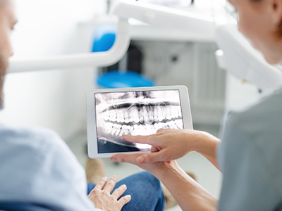
Diagnostic tools such as digital radiography, intraoral photography, and charting of lesion dimensions help document findings and monitor change over time. In many cases the visual and tactile exam provides strong clues about the lesion’s nature. When the diagnosis is uncertain, we discuss the reasons for additional testing and provide a clear plan so patients understand the purpose and potential outcomes.
Part of the clinical visit is patient education: explaining what we see, why it matters, and how we will proceed. We prioritize plain, respectful communication so patients can make informed decisions about biopsies, referrals, or watchful waiting when appropriate.
When a lesion cannot be confidently classified by clinical exam alone, a biopsy is often the next step. Biopsies range from a small surface sample to deeper incisional or excisional procedures, depending on the lesion’s size and suspected nature. These are performed with local anesthesia in a manner designed to minimize discomfort and preserve tissue for accurate analysis.
The tissue specimen is sent to a certified pathology laboratory, where a specialist examines cellular features under the microscope. The pathologist’s report differentiates between inflammatory, infectious, dysplastic (pre-cancerous), and malignant processes. This objective information guides treatment decisions and helps determine whether further imaging, referral to a specialist, or close surveillance is required.
Receiving a pathology result can be stressful, so we help patients interpret findings and outline practical next steps. Whether the report calls for conservative management, surgical removal, or coordination with medical specialists, our goal is a smooth, well-coordinated plan that keeps the patient informed and supported.
Treatment for oral pathology is tailored to the diagnosis and the patient’s overall health. Benign lesions may be observed or removed in-office if they cause symptoms or functional problems. Infectious or inflammatory conditions are treated medically or with targeted interventions. Pre-cancerous changes may require surgical excision or closer clinical surveillance to prevent progression.
For confirmed malignancies, treatment typically involves a multidisciplinary team: oral and maxillofacial surgeons, medical oncologists, radiation oncologists, and other specialists work together to create an individualized care plan. Even when care moves beyond the dental setting, the oral surgery and dental teams remain involved in rehabilitation, oral function, and long-term surveillance to protect the patient’s quality of life.
Because recurrence or new lesions can occur, ongoing monitoring is a crucial part of care. We recommend a follow-up schedule based on the lesion’s nature and the risk profile of the patient, so changes can be detected early and managed promptly. Preventive measures and risk-reduction counseling — including tobacco cessation and safe oral practices — are integrated into long-term follow-up.
Good oral health includes paying attention to changes and seeking evaluation when something is persistent or unusual. Early detection is the most effective strategy for minimizing the impact of oral disease, and a thoughtful diagnostic process reduces unnecessary procedures while ensuring that serious conditions are not overlooked. Patients who notice anything out of the ordinary should not delay scheduling an exam.
Our practice emphasizes clear explanations, careful documentation, and measured decision-making. We prioritize patient comfort and understanding throughout every step — from initial exam to biopsy and follow-up. When collaboration with medical specialists is needed, we facilitate those connections to ensure continuity of care.
If you have questions about a sore, lump, or persistent oral change, contact Advanced Dental Care of Las Colinas for more information and to arrange an evaluation. Early assessment protects oral health and provides peace of mind — we’re here to help you navigate the process with expertise and compassion.
Oral pathology is the study and diagnosis of diseases that affect the mouth, jaws and related structures such as the salivary glands and oral mucosa. It covers a wide spectrum of conditions ranging from benign lesions and infections to precancerous changes and malignancies. Clinicians who evaluate oral pathology look for patterns of tissue change, symptom history and risk factors to determine the likely cause.
Because the mouth reflects both local and systemic health, oral pathology bridges dental and medical care by identifying issues that may require further medical evaluation. Early identification of abnormal findings can lead to less invasive treatments and better long-term outcomes. Regular oral exams are an important part of monitoring for these changes.
Common warning signs include persistent sores or ulcers, unexplained lumps or swellings, white or red patches, numbness, and changes in how teeth fit together when you bite. Some lesions are painless at first, which is why patients and clinicians must pay attention to anything that does not resolve within two weeks. Other symptoms may include persistent pain, difficulty swallowing, or a chronic sore throat.
Symptoms that affect speech, chewing or salivary function should prompt timely evaluation because they can indicate deeper tissue involvement or functional impairment. Not all abnormalities are serious, but documenting and monitoring them helps rule out conditions that require urgent care. Your dentist can track changes over time and recommend diagnostic testing when necessary.
Causes of oral pathology are diverse and include congenital or genetic disorders, infections (viral, bacterial or fungal), traumatic injury, chronic irritation, and environmental exposures such as tobacco or certain chemicals. Some lesions arise from systemic diseases like autoimmune disorders or nutritional deficiencies, while others appear without an identifiable cause and are labeled idiopathic. Lifestyle factors and medication side effects can also produce visible changes in the mouth.
Because many causes overlap, an accurate history and clinical exam are essential to narrow the possibilities. Identifying risk behaviors such as tobacco or heavy alcohol use helps guide both diagnosis and prevention strategies. In some cases, additional medical tests are needed to determine whether an oral finding represents a local problem or part of a broader health issue.
Diagnosis typically begins with a thorough clinical exam and a review of medical history, symptoms and risk factors. Clinicians may use adjunctive tools such as digital radiographs, intraoral photography, special staining, or noninvasive screening devices to better visualize suspicious areas. When visual and clinical findings are inconclusive, a tissue biopsy is often the definitive step to obtain a histopathologic diagnosis.
Laboratory analysis of biopsy samples provides information about the cellular makeup and behavior of a lesion, which guides treatment planning. In some situations, coordination with medical specialists or imaging tests like CT or MRI is necessary to evaluate deeper involvement. Clear communication between dental and medical providers ensures a comprehensive approach to patient care.
An oral biopsy is a minor surgical procedure in which a small sample of tissue is removed for laboratory examination. The area is numbed with local anesthesia so most patients feel little or no pain during the procedure; sensations of pressure are common but typically brief. There are different biopsy techniques, including incisional (sampling a portion) and excisional (removing the entire lesion), and your clinician will choose the approach that best suits the presentation.
Following the biopsy, local bleeding and mild discomfort are expected for a short time and can usually be managed with over-the-counter analgesics and simple aftercare instructions. The site is monitored for proper healing and the pathology report, which may take several days to a couple of weeks, determines the next steps. If results indicate a more serious condition, your dentist will discuss treatment options or arrange a referral to a specialist.
You should seek professional evaluation for any oral lesion that persists longer than two weeks, changes in size or color, causes unexplained pain, or interferes with eating, speaking or swallowing. Immediate attention is warranted for rapidly growing masses, symptoms such as uncontrolled bleeding or increasing numbness, and any lesion in patients with high-risk behaviors like tobacco use. Prompt assessment helps distinguish benign issues from conditions that need urgent treatment.
Routine dental exams are an ideal opportunity to have the mouth screened for abnormalities, even when you have no symptoms. Patients with medical conditions that affect healing or immune function should report new oral findings quickly because they may progress more rapidly. Early detection improves treatment options and outcomes for many oral diseases.
Treatment depends on the underlying diagnosis and can range from conservative medical management to surgical removal. Infections are treated with appropriate antimicrobial therapy, autoimmune-related lesions may need immunomodulatory treatment, and precancerous or cancerous changes often require surgical excision and coordination with oncology specialists. Restorative or rehabilitative care, such as prosthetics or reconstructive surgery, may be part of the treatment plan when tissue removal affects function or appearance.
Follow-up care is essential to monitor healing and detect recurrence or new lesions early. Preventive measures, lifestyle modifications and regular surveillance appointments help reduce the risk of progression for many conditions. Your dental team will explain the rationale for each recommendation and coordinate care with medical colleagues when multidisciplinary treatment is necessary.
Yes — many oral findings reflect systemic health and can be early indicators of diseases such as diabetes, autoimmune disorders, nutritional deficiencies, or infectious conditions. For example, persistent oral ulcers, unusual mucosal changes, or salivary gland enlargement can prompt testing for underlying systemic diseases. Dentists often serve as the first point of detection for conditions that also affect other parts of the body.
When a systemic cause is suspected, the dental clinician may request blood work, imaging, or refer the patient to an appropriate medical specialist for comprehensive evaluation. Collaboration between dental and medical professionals improves diagnostic accuracy and ensures that both oral and overall health needs are addressed. Timely cross-referral helps patients receive the full scope of care they require.
Maintain a consistent oral hygiene routine that includes brushing twice daily with fluoride toothpaste, daily interdental cleaning, and regular professional cleanings. Avoid tobacco in all forms and limit alcohol consumption, as both are significant risk factors for precancerous changes and oral cancer. A balanced diet rich in vitamins and minerals supports mucosal health and overall immune function.
Perform regular self-exams by looking for persistent sores, color changes, lumps or areas of numbness, and report any concerns to your dental provider promptly. Stay current with routine dental visits so your clinician can screen for early signs of disease and provide tailored preventive advice. Vaccination against human papillomavirus (HPV) may also reduce risk for certain types of oral and oropharyngeal cancer; discuss this option with your medical provider.
At Advanced Dental Care of Las Colinas, clinicians begin with a detailed history and thorough oral examination, using diagnostic tools such as digital radiography and clinical photography to document findings. When indicated, we perform or arrange biopsies and send specimens to accredited pathology laboratories for definitive analysis. Our goal is to provide clear explanations of results and a coordinated plan based on the pathology report.
For conditions that require specialist input, the practice maintains relationships with oral surgeons, ENT physicians, oncologists and other medical professionals to ensure timely referrals and collaborative care. We prioritize communication and follow-up so patients understand next steps and receive seamless treatment across providers. If you notice an unusual oral change, our team can evaluate it and guide you through the diagnostic and treatment pathway.
Quick Links